MSK RADIOLOGY
X-rays are types of electromagnetic radiation used to image the skeleton. The test is quick and painless. We are constantly exposed to naturally occurring background radiation with different levels across the country.
X-rays for medical imaging of the limbs and most joints are of low doses, typically equivalent to less than 1.5days of natural background radiation. The dose increases as larger parts of the body are x-rayed and as the x-rays passes through more sensitive organs. Because of this, x-rays must legally be requested by approved practitioners.
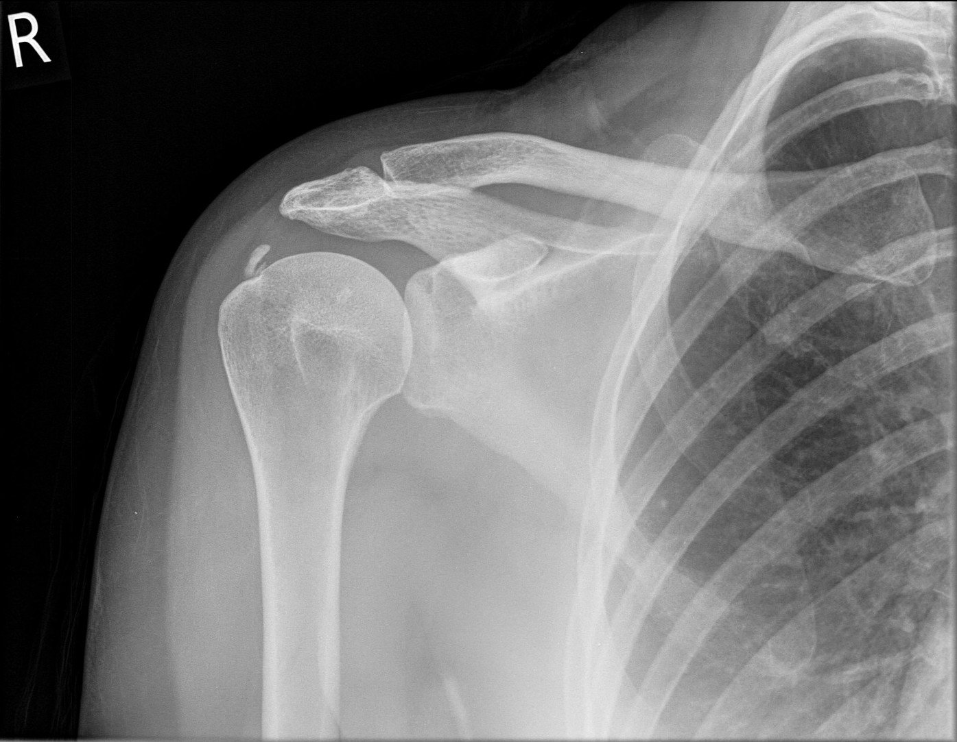
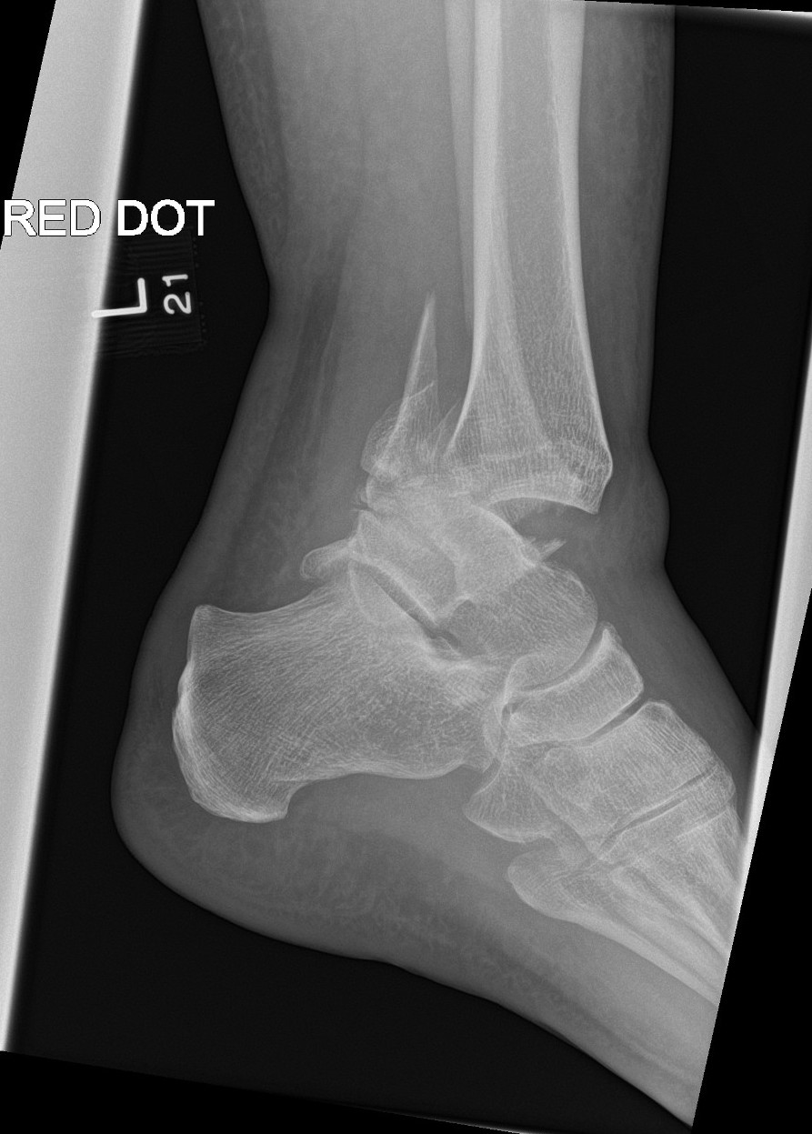
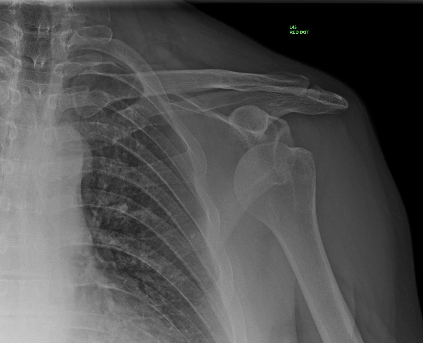
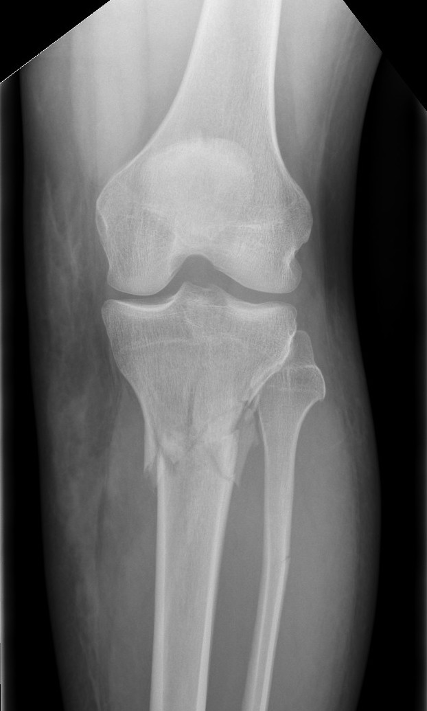
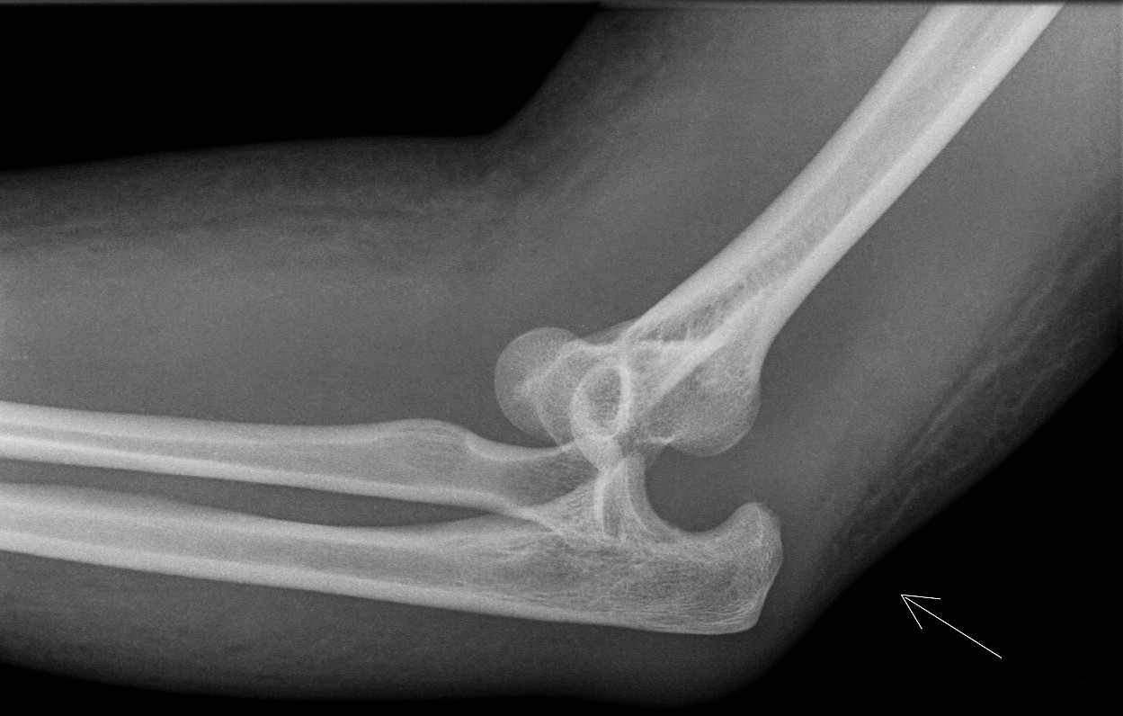
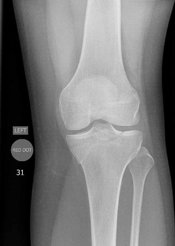
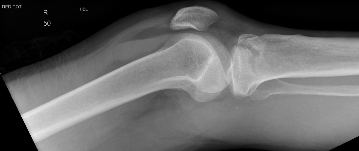
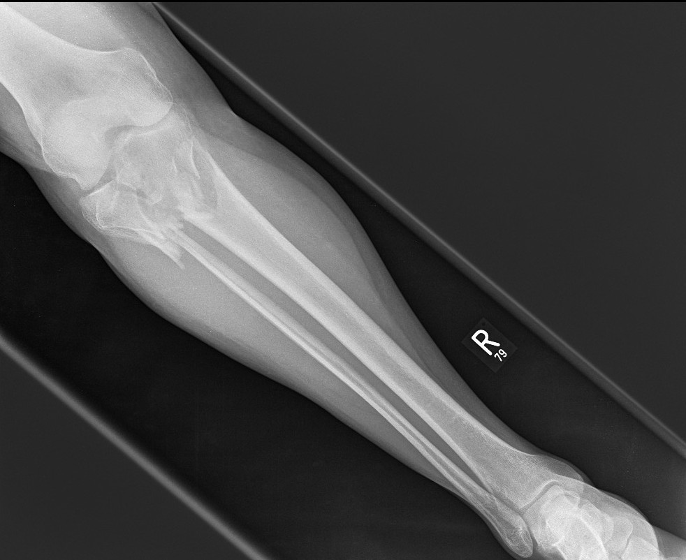
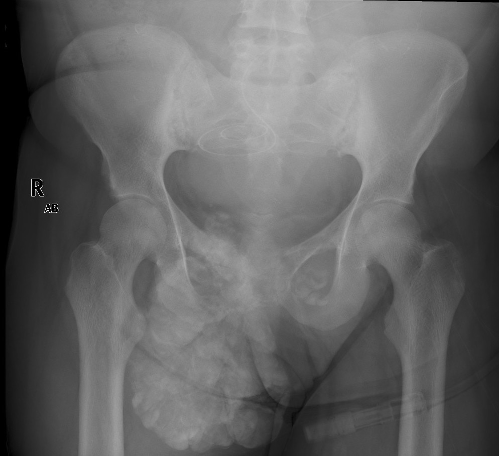
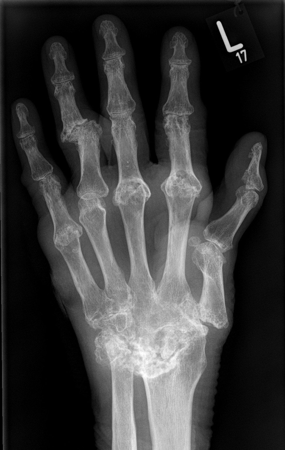
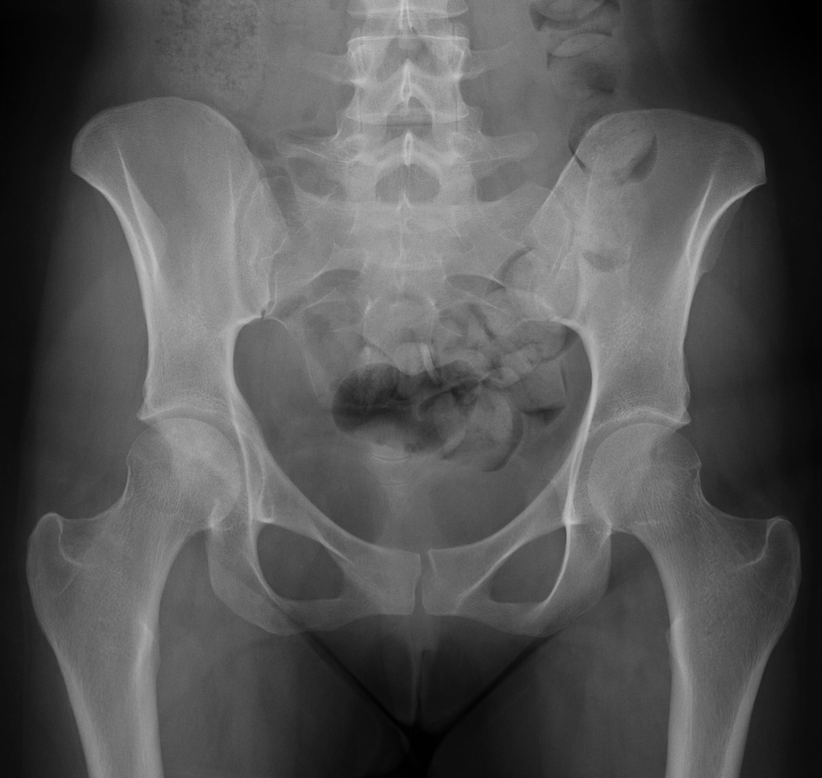
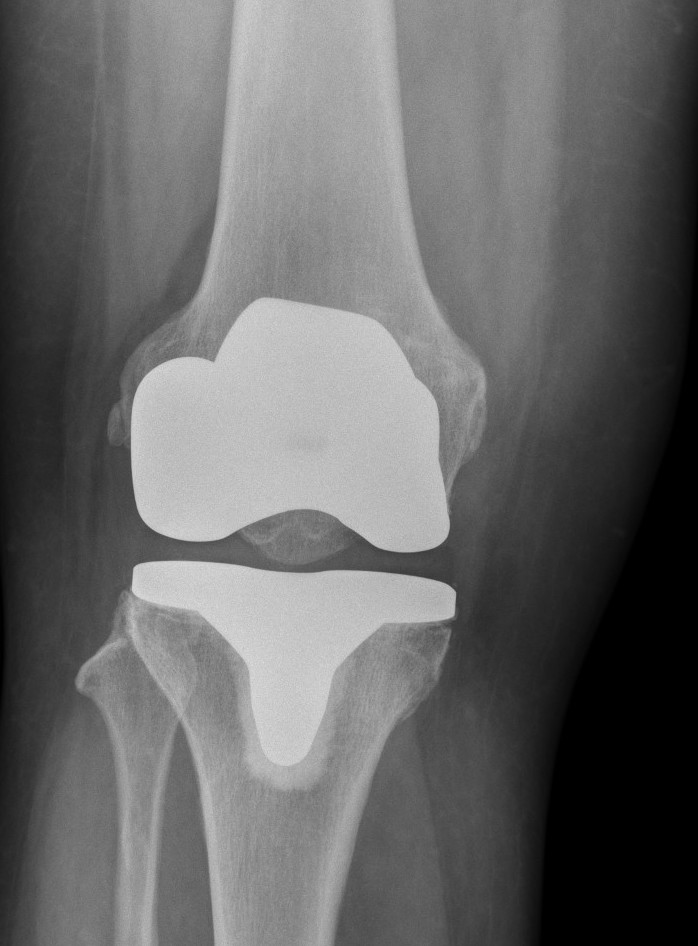
X-rays are extremely useful for detecting problems with bones such as fractures, arthritis and joint replacements.
I read x-rays and provide expert medical opinon for your referring doctor.
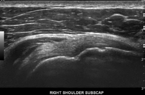
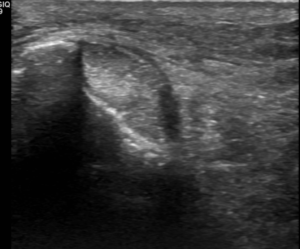
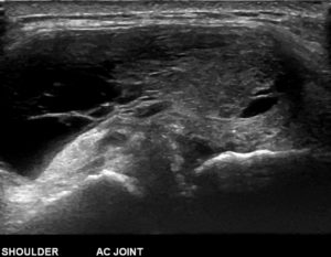
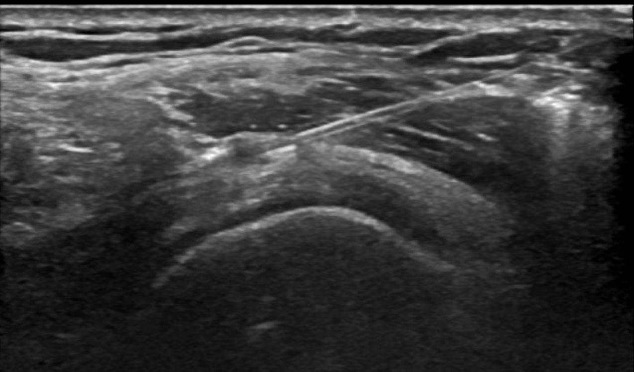
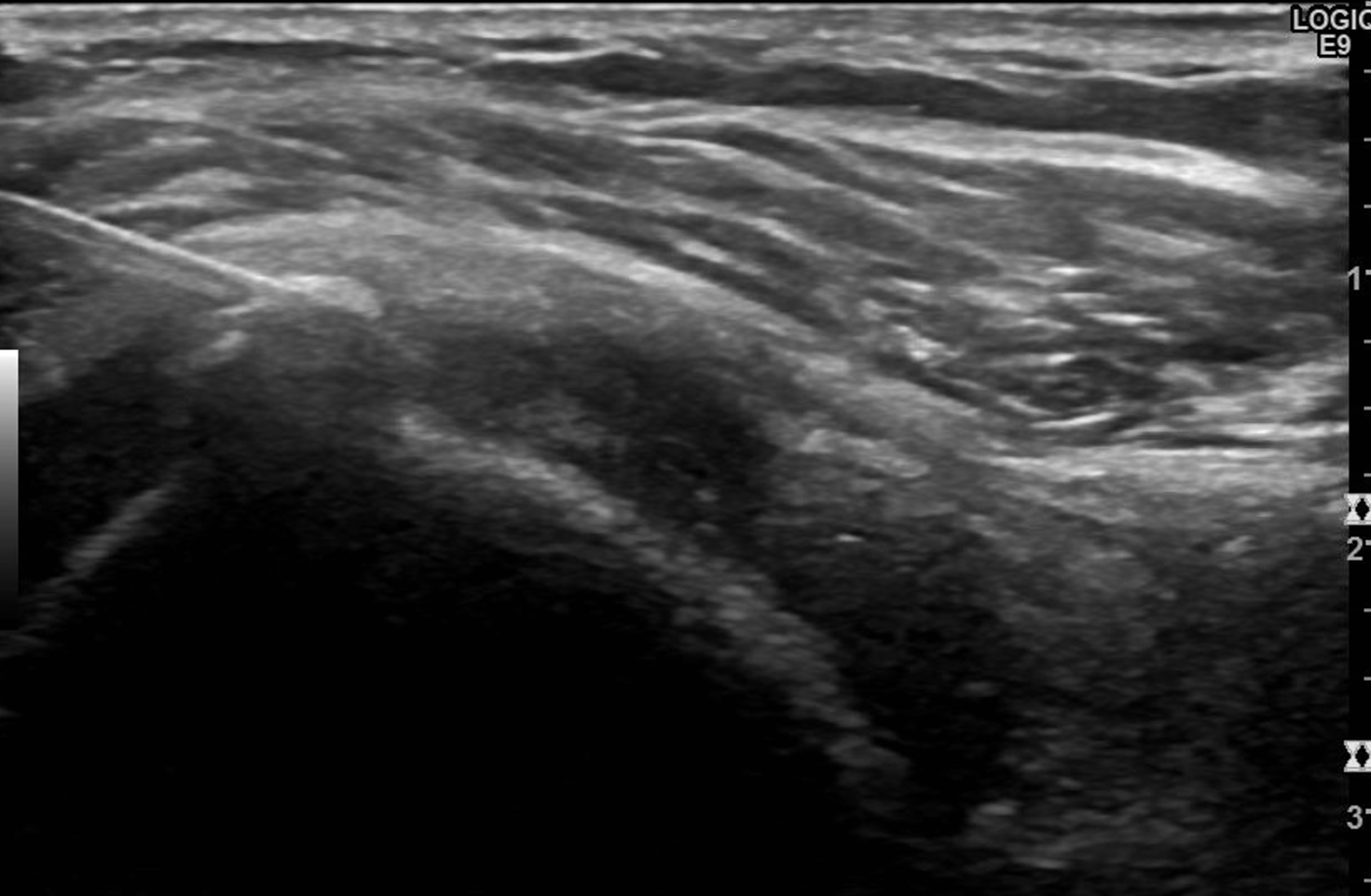
Medical ultrasound uses high frequency sound waves to create images of the internal body. A transducer produces sounds waves which via a gel lubrication, passes through the body and is reflected off internal structures back to the transducer. The ultrasound machine is then able to produce images on the screen. No radiation is involved, is a painless test and is safe for all patients.
Ultrasound is often the first choice of investigation for assessing soft tissues such as muscles and tendons. It is particularly useful to evaluate lumps and bumps. Ultrasound allows me to assess dynamically it is performed in real-time. Ultrasound is limited in its use, in that it cannot assess bones in detail like x-rays or internal structures within a joint.
I will perform a detailed ultrasound of the problematic area and provide an expert medical opinion to your referring doctor.
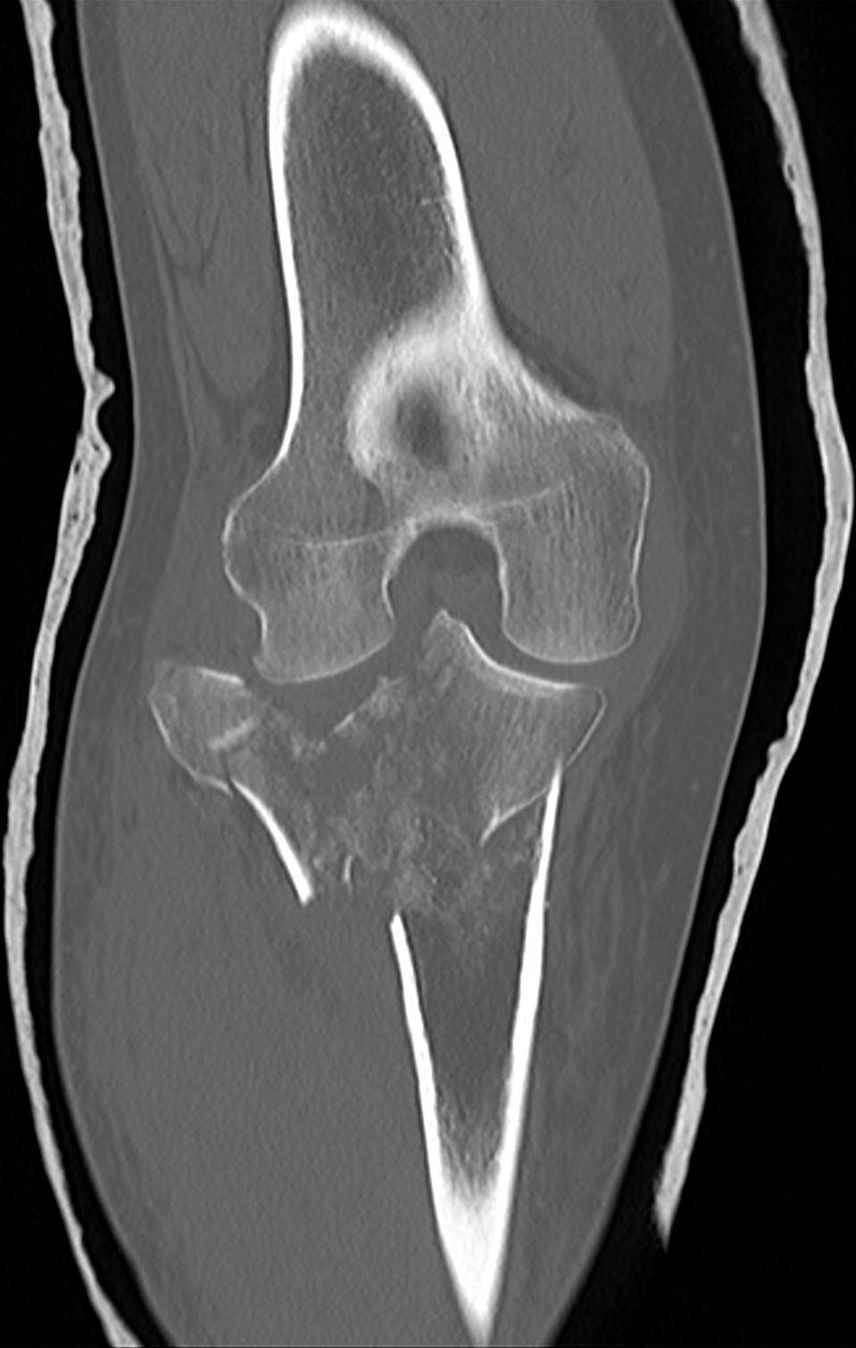
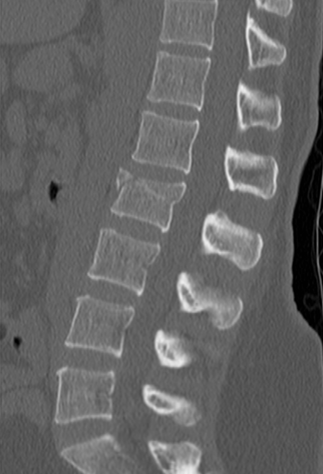
Computer Tomography (CT) uses x-rays within a tube that rotates around you so that a detailed image of the internal body is created at multiple angles. Algorithms within the CT scanner software allows images to be created automatically in multiple planes (axial, coronal and sagittal). The scanner is ring shaped, however, unlike MRI, claustrophobia is rarely reported. The scan time is seconds. Staff must be informed of pregnancy prior to the scan as radiation is used.
CT is extremely useful in providing detailed assessment of the bone for cases of fracture and assessing joints. It is particularly useful for pre-surgical planning to help Orthopaedic Surgeons customise joint replacements.
CTs are performed by radiographers and images are stored to allow me to provide an expert medical opinion to your referring doctor.
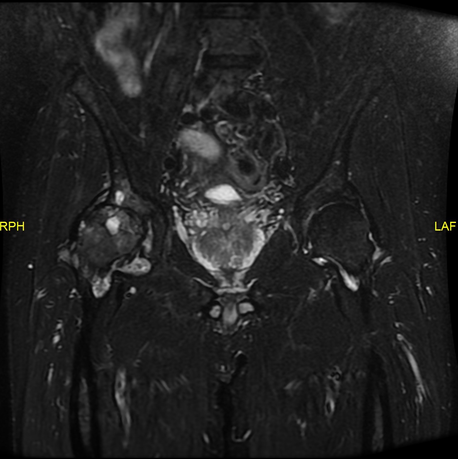
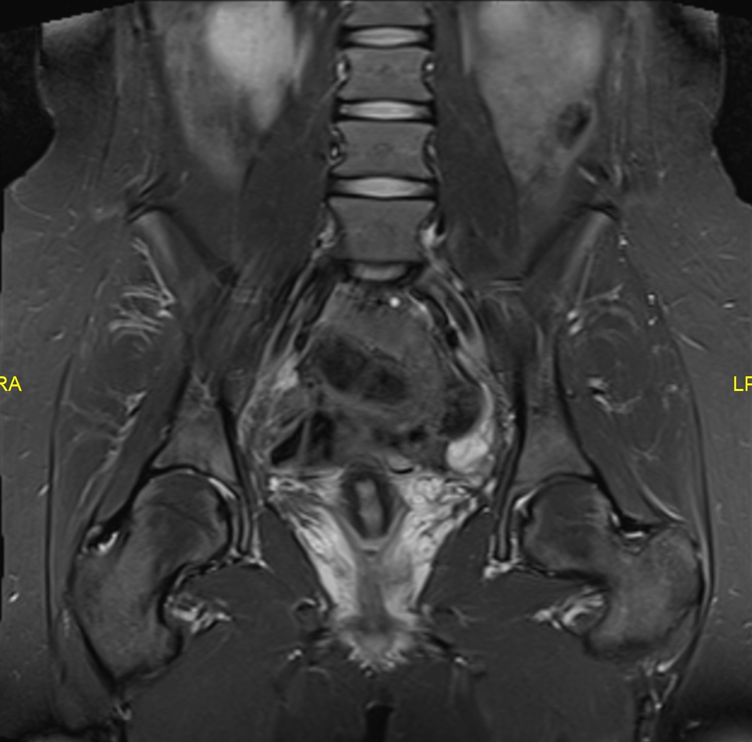
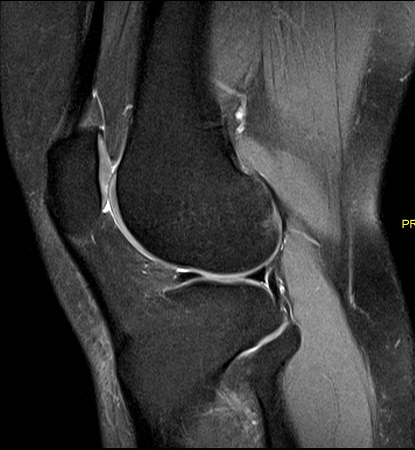
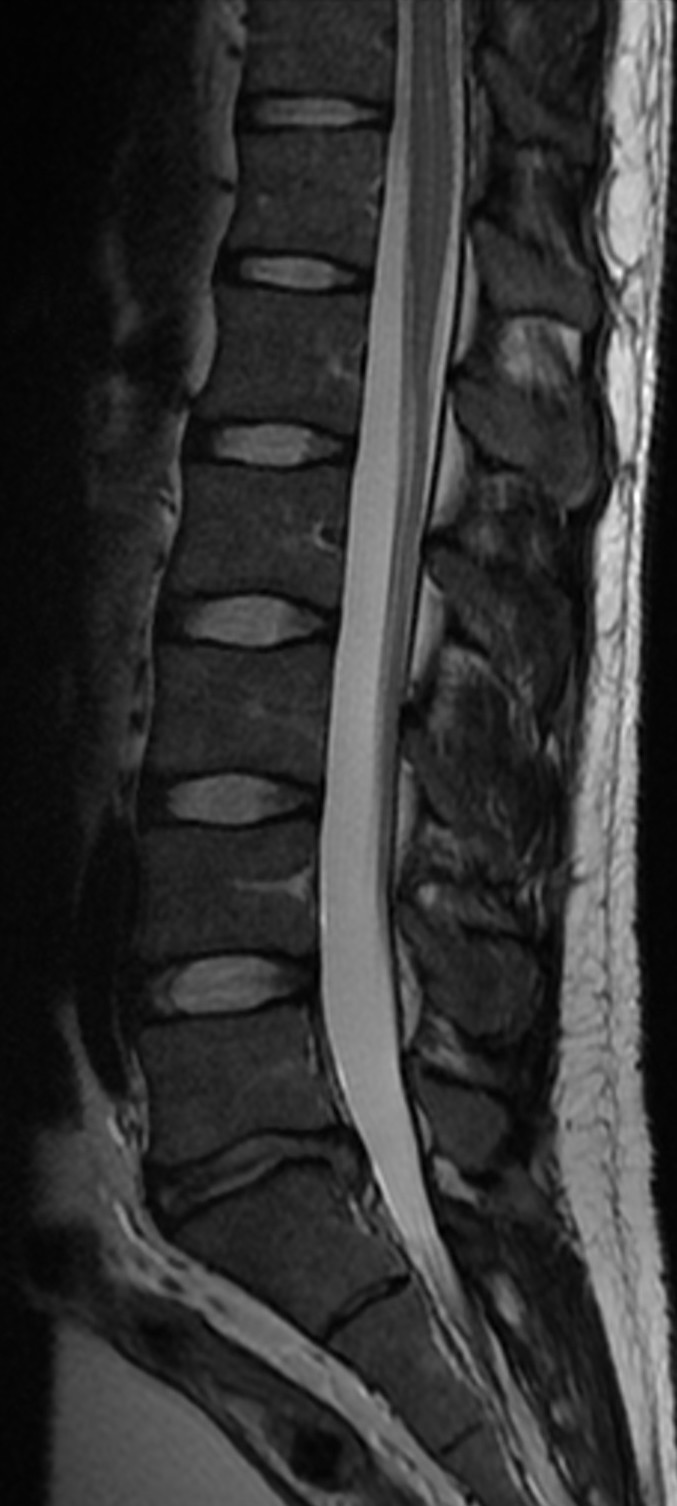
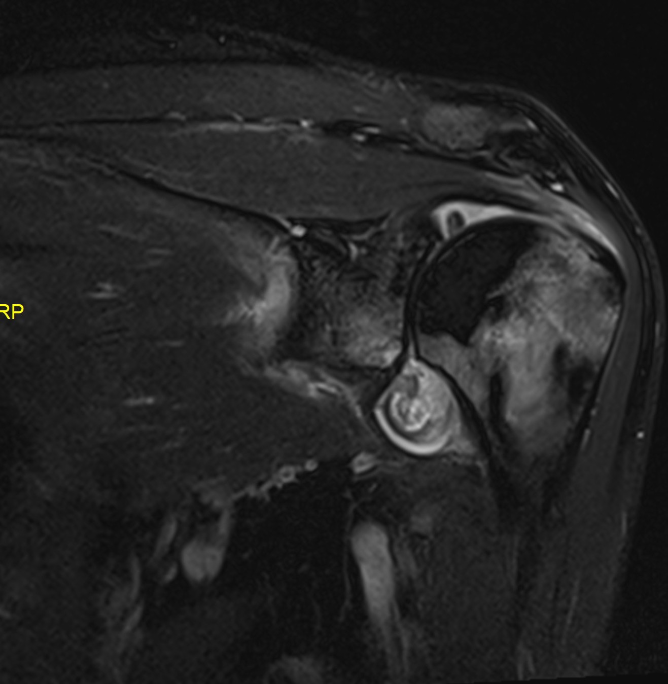
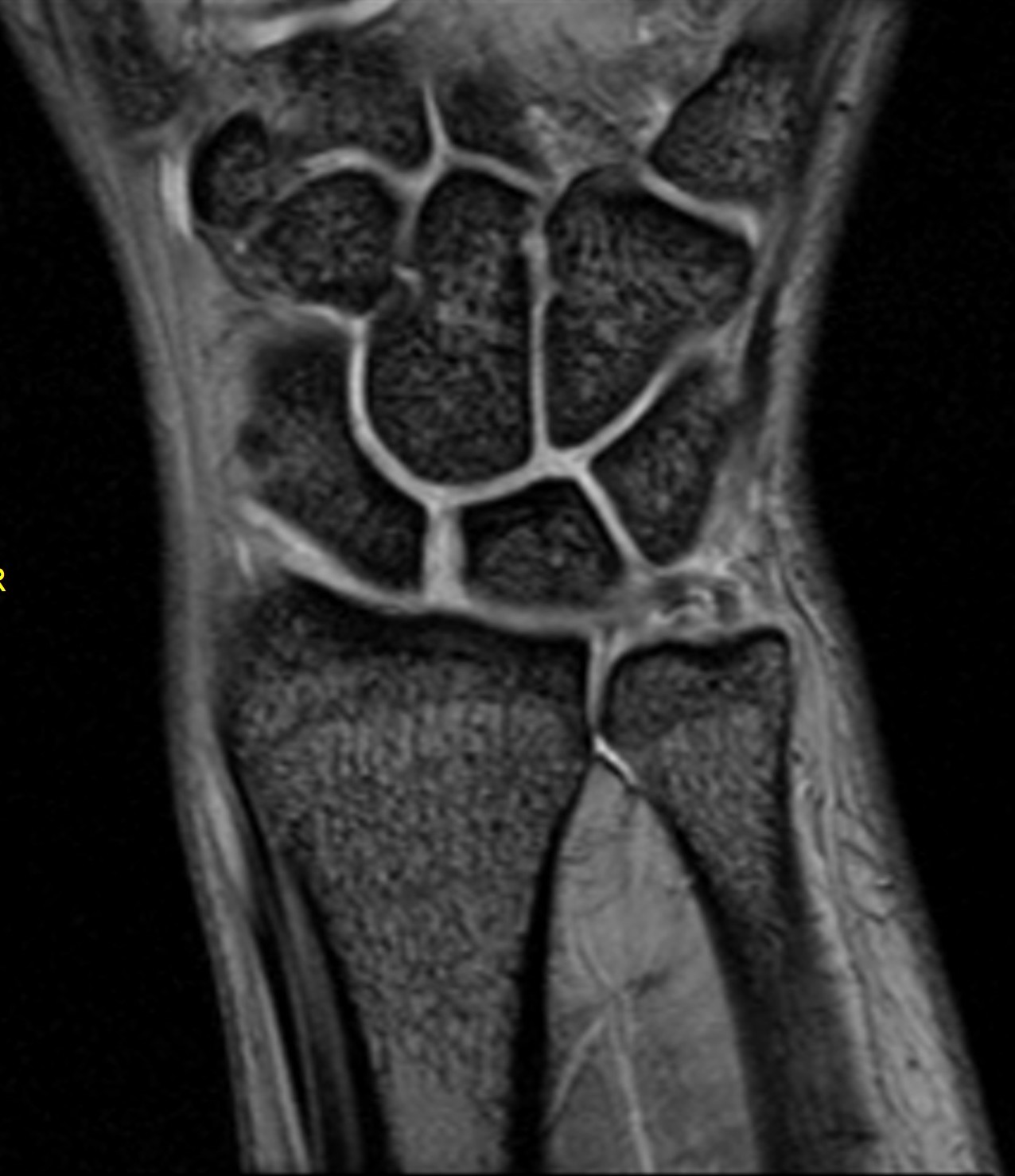
Magnetic Resonance Imaging (MRI) utilises magnetism and radio-waves to produce images without radiation. MRI is exceptionally detailed when evaluating soft tissues like muscles, tendons and ligaments. It allows assessment of small structures within a joint such as cartilage. It allows the assessment of the bone in a different way compared to CT, for example it can assess for bone bruising.
The scanner is ring shaped like a CT scanner, but has a smaller bore which can leave patients feeling claustrophobic. Because of the use of a strong magnet, the scan can be noisy and there are a number of restrictions and precautions one must take before allowing patients near the scanner. MRI may not be suitable for patients with a pacemaker, metal fragments within the body particularly the eyes or metallic cardiac valves. Having a joint replacement (i.e. knee or hip) is safe. Some tattoo inks may contain pigments that are magnetic and thus there have been reported side effects of heating or pulling sensation. MRI can still be performed but may need to be modified.
MRIs are performed by radiographers and images are stored to allow me to provide an expert medical opinion to your referring doctor.
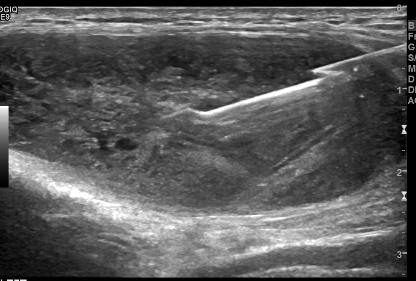
The use of imaging allows the Radiologist to visualise the needle position to ensure precise targeting. The type of imaging used will depend on the type of procedure and area being targeted.
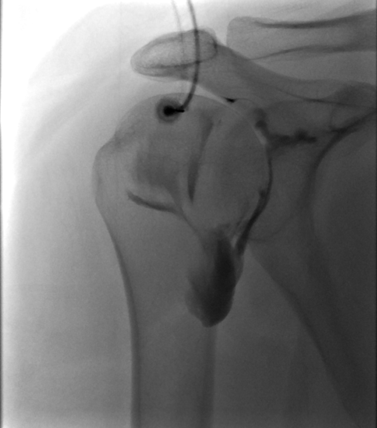
X-rays (or fluoroscopy), ultrasound or CT can be used to perform steroid injections into joints, bursa (fluid sacs) or tendon sheath, aspirate fluid from a collection or biopsy a soft tissue lump or abnormal areas within a bone.
I perform steroid injections to all joints, soft tissue/bursae or tendon sheaths most commonly under ultrasound guidance. This is a safe and comfortable technique with the procedure typically over in seconds. However, appointment times are typically 30mins because of the need to obtain fully informed written consent, perform pre-procedure scans and for sterile preparation of equipment. Ultrasound is also extremely useful for performing biopsies of soft tissue lumps or aspirating fluid e.g. a ganglion in the wrist or a Baker’s cyst in the knee. I also perform more technical procedures such as brisement (Achilles tendon) and barbotage (shoulder).
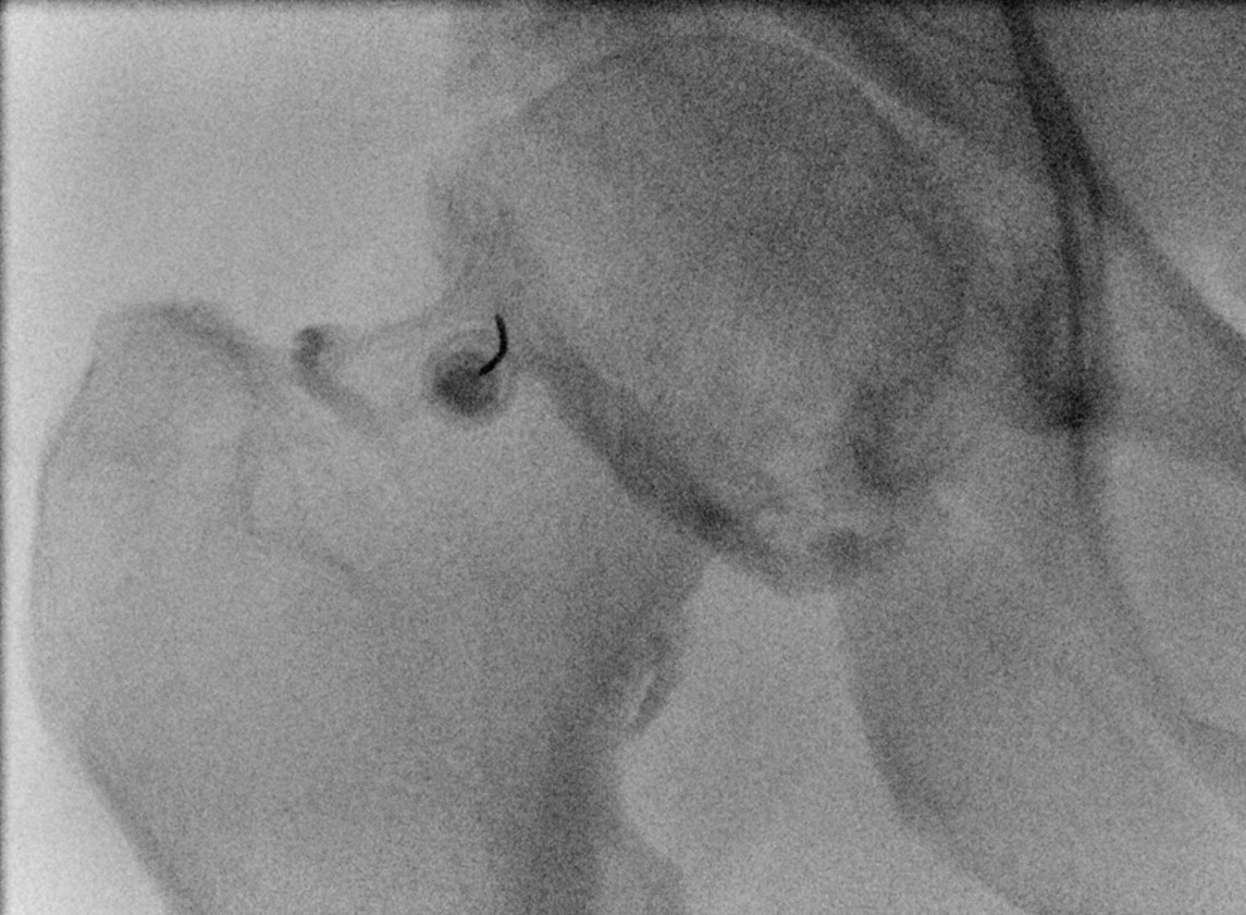
Injections that are required in a deeper location (i.e. shoulder/hip/sacroiliac joint or a nerve root block) often require x-ray guidance using a fluoroscopy machine. This allows dynamic use of real-time low dose x-rays to visualise the necessary anatomy to allow pin-point targeting.
CT can be used for certain spinal injections including facets or nerve root blocks. These can also be performed under fluoroscopy but often depends on the equipment availability which varies at different clinics. If a biopsy of an abnormal area within a bone is required, this will be performed under CT guidance because of the better bone detail.
Most procedures are performed under local anaesthetic (injection or skin spray). Rest assured, I am fully trained and will select the best imaging technique depending on the procedure that allows me to safely perform procedures and that is most suitable for the patient. I have performed hundreds of these procedures over the last 8 years and I perform them on a weekly basis.